

| Title: | Enteroklýza v CT a MR obrazech |
| Other Titles: | Enteroclysis in CT and MR pictures |
| Authors: | Honzíková, Kamila |
| Advisor: | Svobodová, Andrea |
| Referee: | Baxa, Jan |
| Issue Date: | 2012 |
| Publisher: | Západočeská univerzita v Plzni |
| Document type: | bakalářská práce |
| URI: | http://hdl.handle.net/11025/2249 |
| Keywords: | Crohnova choroba;tenké střevo;enteroklýza;CT enterografie;MR enterografie |
| Keywords in different language: | Crohn´s disease;small intestine;enteroclysis;CT enterography;MR enterography |
| Abstract: | Souhrn: Objevením X paprsků panem Wilhelmem Conradem Röntgenem započala éra zobrazovací radiodiagnostiky. Od počátku byly snahy o vyšetřování nejen kostí, ale i dalších orgánů lidského těla. Postupně se stalo i zobrazování tenkého střeva nenahraditelnou součástí radiodiagnostických metod využívaných moderní lékařskou medicínou. Dlouhou dobu měly v algoritmu radiodiagnostických vyšetřovacích metod tenkého střeva výsadní postavení nativní snímek břicha, pasáž GIT a klasická RTG enteroklýza. V druhé polovině 20. století došlo k rozvoji a zdokonalování techniky, kontrastních látek a zejména k rozvoji ultrasonografie, endoskopie, výpočetní tomografie a magnetické rezonance. Cílem mé práce je pomocí teoretické části a kazuistik popsat a porovnat moderní zobrazovací metody CT a MR enterografie, jejich výhody, nevýhody a uplatnění v diagnostice onemocnění tenkého střeva. |
| Abstract in different language: | Summary: The age of imaging radiodiagnostics started with the discovery of x-ray by Wilhelm Conrad Röntgen. Since the very beginning, there were attempts to examine not only the bones but also other human organs. Step by step, viewing the small intestine has become an indispensable part of radiodiagnostics methods used by modern medicine. Native abdominal film, the GIT passage and classical x-ray enteroclysis were for a long time essential in the algorithm of viewing the small intestine. In the second half of the twentieth century, there was technological development, contrast agents and ma-inly there was development of ultrasonography, endoscopy, computal tomography and magnetic resonance imaging. The aim of my work is to describe and compare in the theoretical part and casuistry modern viewing methods of CT and MR enterography with their advantages and disadvantages and how we can use them in diagnosing small intestine illness. |
| Rights: | Plný text práce je přístupný bez omezení. |
| Appears in Collections: | Bakalářské práce / Bachelor´s works (KAZ) |
Files in This Item:
| File | Description | Size | Format | |
|---|---|---|---|---|
| Bakalarska Prace.pdf | Plný text práce | 3,72 MB | Adobe PDF | View/Open |
| Honzikova - VP.jpg | Posudek vedoucího práce | 371,47 kB | JPEG | 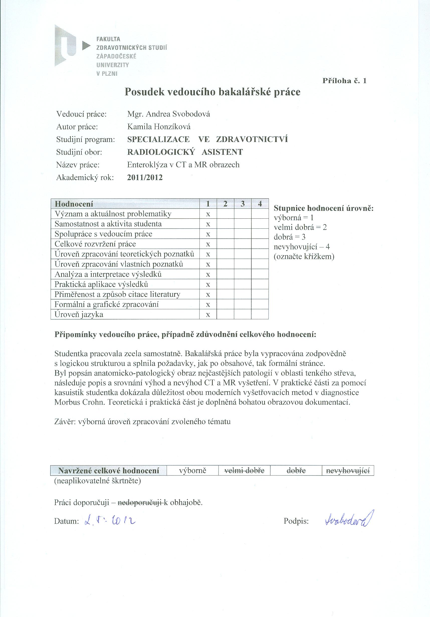 View/Open |
| Honzikova - OP.jpg | Posudek oponenta práce | 332,79 kB | JPEG | 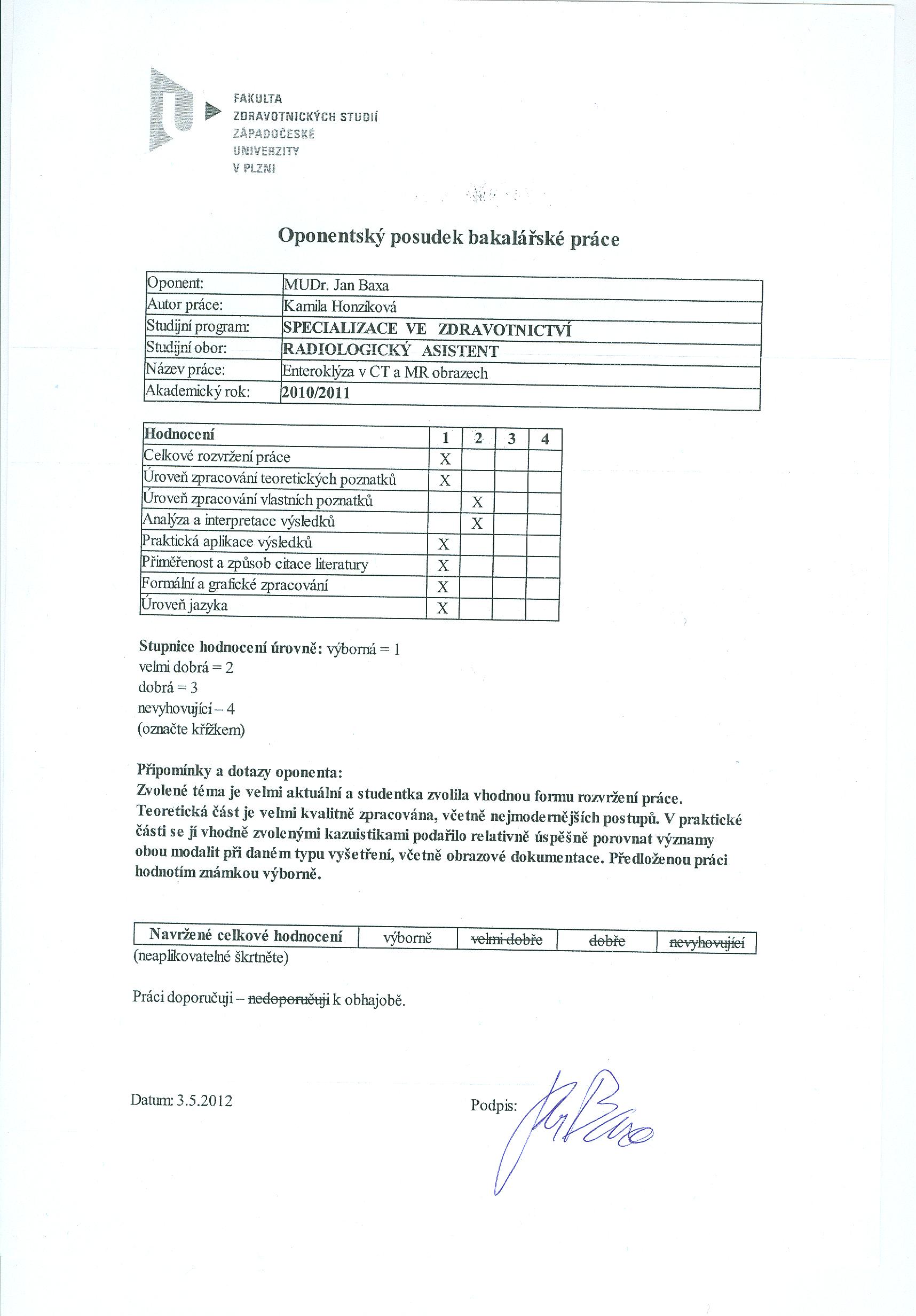 View/Open |
| Honzikova - OBP.jpg | Průběh obhajoby práce | 340,15 kB | JPEG | 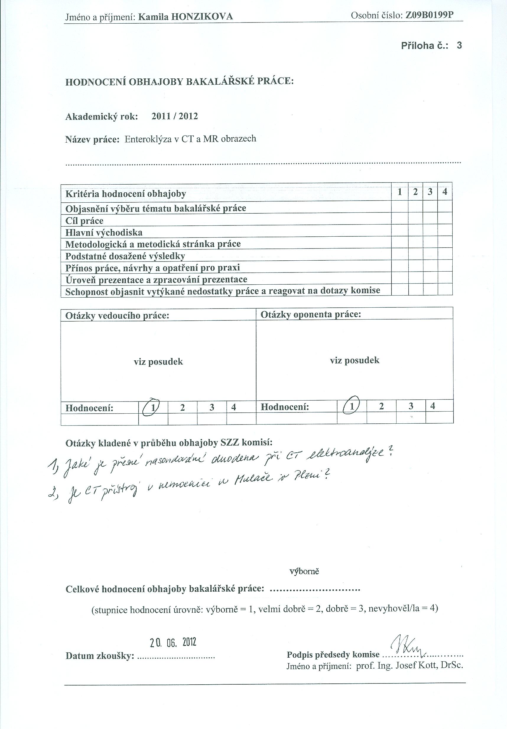 View/Open |
Please use this identifier to cite or link to this item:
http://hdl.handle.net/11025/2249Items in DSpace are protected by copyright, with all rights reserved, unless otherwise indicated.
search
navigation
- DSpace at University of West Bohemia
- Vysokoškolské kvalifikační práce / Theses
- Fakulta zdravotnických studií / Faculty of Health Care Studies
- Katedra záchranářství, diagnostických oborů a veřejného zdravotnictví / Department of Rescue Services, Diagnostic Fields and Public Health
- Bakalářské práce / Bachelor´s works (KAZ)